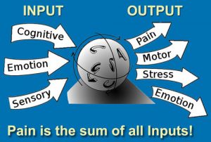
There are several places where the nervous system can be properly tuned, and these “adjusting knobs” are the bones that directly attach to the dura mater. Faulty alignment or fixations in any bone of the cranial vault or spine will over stretch, torsion, malform or drag on the dural membrane, disrupting its ability to send or receive reliable signals from musculoskeletal and visceral structures. These aberrant dural stresses routinely manifest as spasm and pain, and are often misinterpreted as muscle problems. Therapists noticeably benefit from the ability to quickly distinguish between common myofascial pain syndromes and true adverse dural tension signs.
Poor alignment at C2 (axis) can trigger head and neck pain due to the unusual dural membranous attachment to the anterior bodies of the C2-3 joints. Since the notochord is central to the development of the axial skeleton and crucial in determining the final construction of the central nervous system, any distortion here can set-off tonic neck reflexes that travel down intersegmental pathways creating postural havoc in the thorax and pelvis.
All is well so long as the axial skeleton is appropriately aligned and the spinal cord’s protective dural covering is not overstretched by cranial or sacral asymmetries or forward head postures. Sadly, this tough dural tube is commonly distorted by traumatic events or during acts such as prolonged stomach sleeping or poor ergonomic sitting. Whenever the joints of the upper cervical complex (O-A and A-A) and their supporting soft tissues become strained and motion restricted, compensations often travel down the kinetic chain and lock the C2-3 facet joint closed unilaterally. Constant jamming together of the C2-3 joint leads to cartilage derangement which alters the joints axis of rotation, distorts the dural membrane and reciprocally spasms the sensitive suboccipital muscles.
Most of us refer to the ‘deep eight’ suboccipitals as muscles … and that’s OK. A number of researchers believe their primary function is that of proprioception i.e., helping the vestibular system interpret where the head is in space. Rather, as Ida Rolf would say: “Suboccipitals tell the brain which end is up!” Since the suboccipitals consist of Golgi end organs instead of Golgi tendon organs (as they attach to the skull), by definition, they should be classified as ligaments. Exactly why do they contain end-organs instead of tendon organs? Mother Nature held a grand design here.
Check out my instructional video: http://youtu.be/l6aOWLBFWqI
On sale this week only!
Save 25% off the Posture Pain Performance course!

NEW! USB version with enhanced video
Discover the foundational principles behind MAT technique as we take you on an in-depth look at the connection between pain, posture and function. Save 25% off the Posture Pain Performance course this week only. Offer expires Monday April 22nd. Click the button below for more information and to purchase the course. Upon completion receive 20CE hours and a certificate of completion to display in your office.
Bonus: Order the Home Study version and receive the e-Course for FREE!